Magnetic resonance imaging (MRI) may be used to examine your spinal cord for injuries, the presence of structural abnormalities, or other certain conditions to see if you’re the right candidate for outpatient spine surgery.
Some of these conditions include disc and joint disease, spine misalignment, birth defects in the vertebrae or spinal cord, compression or inflammation of the spinal cord and nerves, trauma injury to the bone, disc, ligament or spinal cord, and herniation or degeneration of discs.
MRI: What is It? And Why Outpatient Spine Surgery?
You can get an MRI through outpatient services or in a hospital. Your doctor’s practice and your condition may determine the procedure, especially if it’s being used to determine next-steps for back pain treatment, therapy, or outpatient spine surgery.
You’ll need to take off any clothing, jewelry, eyeglasses, hearing aids, hairpins, removable dental work, and other objects that could interfere. A gown will be given to you if you need to take off your clothes. To inject contrast dye, an intravenous line (IV) will be gently attached to your hand or arm.
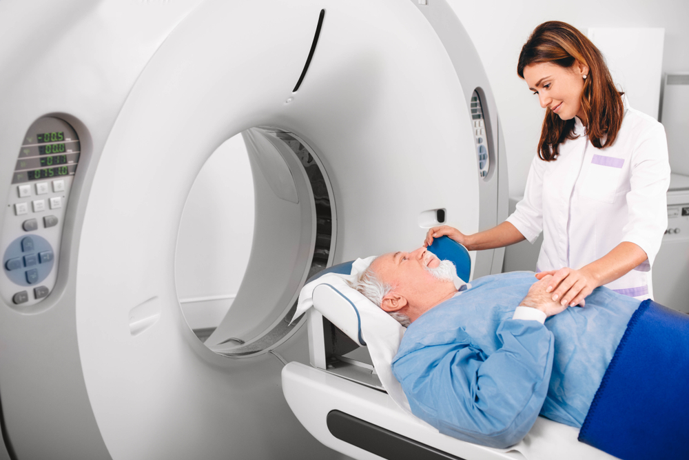
In the scanning machine, you’ll lie on a table that slides into a circular opening. You can use pillows and straps to keep still during the procedure. It will be the specialist’s job to control the scanner in another room. You’ll always be able to see the specialist through a window. You can hear and talk to the specialist through speakers inside the scanner, and that expert will have a call button ready for you in case you have any issues. Being in constant and convenient communication with the specialist, you’ll be monitored safely and securely.
To block out the scanner’s noise, you’ll be given earplugs or a headset. You can usually listen to music if you want. You’ll hear a clicking noise as the magnetic field is created and radio waves are sent from the scanner.
During the scan, you should stay very still, because any movement can distort it. The body part being examined may require you to hold your breath or not breathe for a few seconds at a time. You should not have to hold your breath for longer than a few seconds, which makes the process even easier.
When contrast dye is injected into the IV line, you might feel some effects. A light flushing sensation or feeling of coldness is sometimes evident, or even a metallic or salty taste in your mouth for a short period of time.
You’ll be gently and comfortably assisted off the table once the scan is done. MRIs are painless, but lying still for the whole procedure might take some grit. Just relax and stay calm — it’s the best and most contemplative way to go through the procedure. Many patients prepare themselves by softly meditating and relaxing in the time (or day) before the MRI scan. To minimize any discomfort, the specialist will use all possible measures available. Your comfort is of utmost importance throughout the entire process.
Outpatient Spine Surgery: After the MRI, What Happens?
It’s best to move slowly when getting up from the scanner table to avoid dizziness or lightheadedness. You might need to rest after the procedure if you took any sedatives. Driving will also be a no-no.
It’s possible to be monitored for side effects or reactions to the contrast dye during your procedure, like itching, swelling, rash, or something else. However, after your procedure, if you notice any pain, redness, or swelling at the IV site, you should tell your doctor.
MRIs of the spine don’t need special care. Unless your doctor says otherwise, you can resume your usual diet and activities. Yet, depending on your situation, your doctor might give you additional instructions after the procedure.
You will soon know afterward what the MRI found with respect to your back pain, lack of mobility, or any other issues causing you trouble. It will inform you whether you’ll want to consider outpatient spine surgery.
So How Did the MRI Scan Work?
The scanning machine is a big, cylindrical tube-shaped modern marvel that creates a strong magnetic field around the patient. Radio waves and magnetic fields change hydrogen atoms’ alignment in the body. Then, a scanner sends radio waves that knock the atoms’ nuclei out of place. When the nuclei realign, they send out radio signals. Computers analyze and convert these signals into two-dimensional (2D) images of body structures or organs.
If you’re looking at organs or soft tissue, an MRI is better than a computed tomography scan (CT scan) because it can tell the difference between normal and abnormal tissues.
Additional magnetic resonance technology has been developed because of new uses and indications. In fact, MRA (magnetic resonance angiography) is a non-invasive way to check blood flow through arteries. In addition to detecting aneurysms (such as inside the brain), an MRA can also detect vascular malformations (abnormalities of blood vessels in the brain).
Also, magnetic resonance spectroscopy (MRS) is another non-invasive way to check for chemical changes in body tissues. You can use MRS to diagnose certain infections, strokes, head injuries, comas, Alzheimer’s, tumors, and multiple sclerosis.
A functional magnetic resonance imaging scan (fMRI) determines where in the brain a certain function happens, like speech or memory. There are general brain areas where such functions happen, but the exact location may vary from person to person. Doctors can pinpoint the exact location of the functional center in the brain, and then they can plan surgery or other treatments.
Having outpatient spine surgery is a big decision, and having an MRI is just one step in the entire process. But with today’s “awake” spinal surgical technology and options — along with local anesthesia, which is much better for most patients — you’ll be in the safest of hands.
Spine Science & MRIs: Educate Yourself
To understand more about the anatomy of your back, who gets back pain, the types of back pain, symptoms, and causes, you need to understand the basics of your spine and back pain altogether.
There are 24 vertebrae (individual vertebrae) in the spinal column, plus two naturally fused vertebrae at the bottom called the sacrum and the coccyx. People usually mean the vertebral column when they talk about the spinal column (those 24 circular vertebrae running down the middle). There are five regions of the vertebral column, too.
Here, we explain the basics about your spinal anatomy to help you understand your back or neck pain, as well as a spine surgeon’s potential diagnosis and outpatient spinal surgery or spinal fusion treatment plan. Learn about the five areas of your spinal column, including your sacrum, coccyx, ligaments, discs, facet joints, muscles, spinal nerves, vertebral column, spaces, coverings, and much more.
Sometimes, someone’s back pain is so intense that even a naturally born strong vertebral column may need outpatient spine surgery. That’s where a spinal surgeon’s examination and recommendations can be very helpful.
Awake Spinal Fusion is here to educate you on the basics of your spine, back pain, “awake” spinal and neck surgery, minimally invasive surgery options, and much more.


