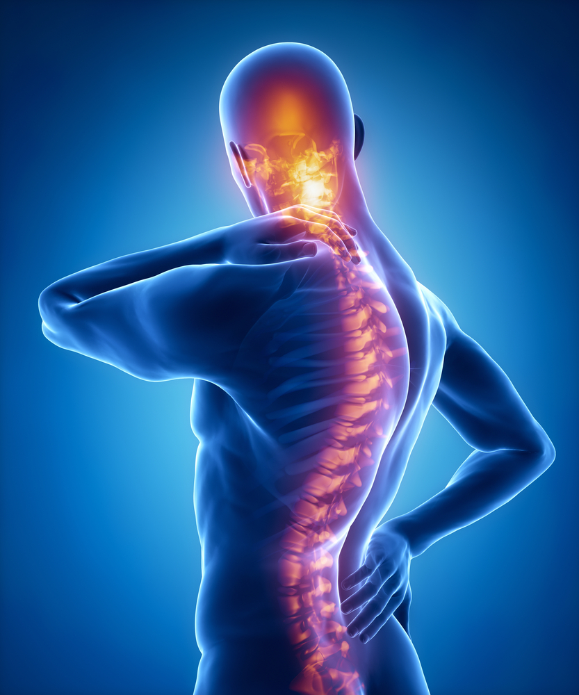
To understand more about the anatomy of your back, who gets back pain, the types of back pain, symptoms, and causes, you need to understand the basics of your spine and the basics of back pain altogether.
“Low back pain is a very common complaint for a simple reason,” states one reputable source. “Since the lumbar spine is connected to your pelvis, this is where most of your weight-bearing and body movement takes place. Typically, this is where people tend to place too much pressure, such as lifting a heavy box, twisting to move a heavy load, or carrying a heavy object. Such repetitive injuries can lead to damage to the parts of the lumbar spine.”
Here, we explain the basics about your spinal anatomy to help you understand your back or neck pain, as well as a spine surgeon’s potential diagnosis and outpatient spinal surgery or spinal fusion treatment plan.
Five Areas of Your Spinal Column
All of your body’s structure and nervous system are supported by the spinal column (vertebral column). There are 34 bones in the spinal column, which holds the body upright, lets it bend and twist easily, and transmits nerve signals from your brain to your toes.
The spinal column is made up of these parts:
- Cervical spine, including the diaphragm (breathing), shoulders, arms, esophagus, and part of the chest.
- Thoracic spine, including parts of the esophagus and arm, trachea, heart, lungs, liver, gallbladder, and small intestine.
- Lumbar spine: the legs and feet.
- Sacrum: bowels, bladders, sexual organs (function and activity).
Your Spinal Column: Think ‘Bone Scaffolding’
There are 24 vertebrae (individual vertebrae) in the spinal column, plus two naturally fused vertebrae at the bottom called the sacrum and the coccyx. People usually mean the vertebral column when they talk about the spinal column (those 24 circular vertebrae running down the middle).
There are five regions of the vertebral column:
- Sacrum (shield-shaped bony structure that is located at the base of the lumbar vertebrae and connected to the pelvis).
- Coccyx (a small triangular bone at the base of the spinal column in humans and some apes, formed of fused vestigial vertebrae).
- Lumbar spine: five vertebrae of the lower back (L1 – L5).
- Thoracic spine: 12 vertebrae of the mid-back (T1 – T12).
- Cervical spine: seven vertebrae of the neck (C1 – C7).
- C1 is the atlas (allowing rotation of the head, as well as extension and flexibility of the head).
- C2 is the axis (forms a pivot-point on which the C1 atlas can rotate).
Viewed from the side, the vertebral column has a graceful double-S curve. In the cervical spine, the vertebrae gently curve inward. In the thoracic spine, the vertebrae gently curve outward. In the lumbar spine, they curve inward again. As a result, the spinal column is strong and shock-absorbing.
Sometimes, though, someone’s back pain is so intense that even a naturally-born strong vertebral column may need non-invasive back surgery. That’s where a spinal surgeon’s examination and recommendations can be very helpful.
Your Sacrum and the Coccyx
Sacrums are triangular-shaped bones located below the last lumbar vertebrae. The sacrum is the backbone of the pelvis and sits between the hip bones. The sacroiliac joints (SI joints) connect the sacrum to the pelvis on both sides.
The coccyx (or tailbone) is formed when three to five small bones naturally fuse together at adulthood. The coccyx is sometimes called the coccygeal vertebrae. When you sit, the tailbone supports your weight, even though it’s small and may seem insignificant.
Ligaments, Discs, Facet Joints and Muscles
Bones aren’t the only thing that makes up the spinal column. The spine also relies on a bunch of supporting structures to maintain its shape, support the skeleton, and route nerves.
Intervertebral discs are the first of these structures. The discs are made up of fibrous tissue (fibrocartilage) and are attached to the vertebrae by cartilaginous endplates, starting at C3 and going all the way to L5. They act as shock absorbers and spacers between the bones. Neither the sacrum nor the coccyx have spinal discs between C1 and C2.
Each vertebral body (C3 – L5) has a pair of facet joints (left and right). Sometimes, individually, they’re called zygapophyseal or apophysis. Besides stabilizing the spine, these joints let you bend forward, extend backward, and twist (called articulation). In the same way as other joints in the body, the facet joint is surrounded by a capsule of connective tissue that produces lubricating fluid. Joint surfaces are coated in cartilage, so they move smoothly.
The spinal column is supported by strong, tough ligaments that connect the vertebrae, discs, and facet joints. A ligament acts like a stretchy tension cord that lets bones, discs, and joints move within a limited range.
The vertebral column is also stabilized and strengthened by small and large spinal muscles and tendons while supporting and restricting extreme bending, flexing, and twisting. For those needing it, outpatient spinal surgery is oftentimes the answer to major back issues affecting the vertebral column.
Spinal Nerves: Protected by the Vertebral Column
Spinal cords and nerve roots are protected by the vertebral column, which is part of the central nervous system. Your spinal cord is housed in a round hollow space between the vertebral structures.
Neuroforamen are openings in the vertebral column where small nerve roots exit the spinal cord. Intervertebral discs are anchored between two vertebrae, and each has a foraminal space. Peripheral nerves branch out from the spinal column and serve the entire body.
The spinal cord ends at L1, and the sacrum and becomes a fan-shaped network of nerves called the cauda equina.
Your Interweaving Nerves and the Vertebral Column
You can think of the foramen like highway exits on the spinal canal. Normally, spinal nerves exit the spinal canal through the openings associated with the part of the body they support. Nerves enable movement and sensation. Since the cervical spine is closest to the hands, nerves that provide sensation to the fingers exit the spinal canal through these openings.
Specialists in spine care know everything about the nervous system, including its causes and effects. An examination of the patient’s physical and neurological symptoms helps spine doctors diagnose problems, such as whether pain is a small sciatica problem versus a larger herniated disc issue. If you ever research today’s safe spine surgery techniques and procedures, you’ll find that the entire field has come a long, long ways in research and innovations.
Spaces and Coverings
Meninges cover the spinal cord (three layers of membranes). Pia mater is intimately connected to the cord. Arachnoid mater is the next membrane. And then there’s a tough outer membrane called the dura mater.
Diagnostic and treatment procedures take place between these membranes. Subarachnoid space surrounds the spinal cord and contains cerebrospinal fluid (CSF) between pia and arachnoid mater. Most often, this space is accessed during lumbar punctures to sample fluid. There’s a space between the dura mater and the bone called the epidural space. It’s usually used for anesthetics, commonly called epidurals, and steroid injections.
Get Informed — and Examined
To know what’s going on with your back pain, you’ve got to know your back first. anatomy of your back, who gets back pain, the types of back pain, symptoms, and causes, you need to understand the basics of your spine and the basics of back pain altogether.
“Low back pain is a very common complaint for a simple reason,” states one reputable source. “Since the lumbar spine is connected to your pelvis, this is where most of your weight-bearing and body movement takes place. Typically, this is where people tend to place too much pressure, such as lifting a heavy box, twisting to move a heavy load, or carrying a heavy object. Such repetitive injuries can lead to damage to the parts of the lumbar spine.”
Here, we explain the basics about your spinal anatomy to help you understand your back or neck pain, as well as a spine surgeon’s potential diagnosis and outpatient spinal surgery or spinal fusion treatment plan.
Five Areas of Your Spinal Column
All of your body’s structure and nervous system are supported by the spinal column (vertebral column). There are 34 bones in the spinal column, which holds the body upright, lets it bend and twist easily, and transmits nerve signals from your brain to your toes.
The spinal column is made up of these parts:
- Cervical spine, including the diaphragm (breathing), shoulders, arms, esophagus, and part of the chest.
- Thoracic spine, including parts of the esophagus and arm, trachea, heart, lungs, liver, gallbladder, and small intestine.
- Lumbar spine: the legs and feet.
- Sacrum: bowels, bladders, sexual organs (function and activity).
Your Spinal Column: Think ‘Bone Scaffolding’
There are 24 vertebrae (individual vertebrae) in the spinal column, plus two naturally fused vertebrae at the bottom called the sacrum and the coccyx. People usually mean the vertebral column when they talk about the spinal column (those 24 circular vertebrae running down the middle).
There are five regions of the vertebral column:
- Sacrum (shield-shaped bony structure that is located at the base of the lumbar vertebrae and connected to the pelvis).
- Coccyx (a small triangular bone at the base of the spinal column in humans and some apes, formed of fused vestigial vertebrae).
- Lumbar spine: five vertebrae of the lower back (L1 – L5).
- Thoracic spine: 12 vertebrae of the mid-back (T1 – T12).
- Cervical spine: seven vertebrae of the neck (C1 – C7).
- C1 is the atlas (allowing rotation of the head, as well as extension and flexibility of the head).
- C2 is the axis (forms a pivot-point on which the C1 atlas can rotate).
Viewed from the side, the vertebral column has a graceful double-S curve. In the cervical spine, the vertebrae gently curve inward. In the thoracic spine, the vertebrae gently curve outward. In the lumbar spine, they curve inward again. As a result, the spinal column is strong and shock-absorbing.
Sometimes, though, someone’s back pain is so intense that even a naturally-born strong vertebral column may need non-invasive back surgery. That’s where a spinal surgeon’s examination and recommendations can be very helpful.
Your Sacrum and the Coccyx
Sacrums are triangular-shaped bones located below the last lumbar vertebrae. The sacrum is the backbone of the pelvis and sits between the hip bones. The sacroiliac joints (SI joints) connect the sacrum to the pelvis on both sides.
The coccyx (or tailbone) is formed when three to five small bones naturally fuse together at adulthood. The coccyx is sometimes called the coccygeal vertebrae. When you sit, the tailbone supports your weight, even though it’s small and may seem insignificant.
Ligaments, Discs, Facet Joints and Muscles
Bones aren’t the only thing that makes up the spinal column. The spine also relies on a bunch of supporting structures to maintain its shape, support the skeleton, and route nerves.
Intervertebral discs are the first of these structures. The discs are made up of fibrous tissue (fibrocartilage) and are attached to the vertebrae by cartilaginous endplates, starting at C3 and going all the way to L5. They act as shock absorbers and spacers between the bones. Neither the sacrum nor the coccyx have spinal discs between C1 and C2.
Each vertebral body (C3 – L5) has a pair of facet joints (left and right). Sometimes, individually, they’re called zygapophyseal or apophysis. Besides stabilizing the spine, these joints let you bend forward, extend backward, and twist (called articulation). In the same way as other joints in the body, the facet joint is surrounded by a capsule of connective tissue that produces lubricating fluid. Joint surfaces are coated in cartilage, so they move smoothly.
The spinal column is supported by strong, tough ligaments that connect the vertebrae, discs, and facet joints. A ligament acts like a stretchy tension cord that lets bones, discs, and joints move within a limited range.
The vertebral column is also stabilized and strengthened by small and large spinal muscles and tendons while supporting and restricting extreme bending, flexing, and twisting. For those needing it, outpatient spinal surgery is oftentimes the answer to major back issues affecting the vertebral column.
Spinal Nerves: Protected by the Vertebral Column
Spinal cords and nerve roots are protected by the vertebral column, which is part of the central nervous system. Your spinal cord is housed in a round hollow space between the vertebral structures.
Neuroforamen are openings in the vertebral column where small nerve roots exit the spinal cord. Intervertebral discs are anchored between two vertebrae, and each has a foraminal space. Peripheral nerves branch out from the spinal column and serve the entire body.
The spinal cord ends at L1, and the sacrum and becomes a fan-shaped network of nerves called the cauda equina.
Your Interweaving Nerves and the Vertebral Column
You can think of the foramen like highway exits on the spinal canal. Normally, spinal nerves exit the spinal canal through the openings associated with the part of the body they support. Nerves enable movement and sensation. Since the cervical spine is closest to the hands, nerves that provide sensation to the fingers exit the spinal canal through these openings.
Specialists in spine care know everything about the nervous system, including its causes and effects. An examination of the patient’s physical and neurological symptoms helps spine doctors diagnose problems, such as whether pain is a small sciatica problem versus a larger herniated disc issue. If you ever research today’s safe spine surgery techniques and procedures, you’ll find that the entire field has come a long, long ways in research and innovations.
Spaces and Coverings
Meninges cover the spinal cord (three layers of membranes). Pia mater is intimately connected to the cord. Arachnoid mater is the next membrane. And then there’s a tough outer membrane called the dura mater.
Diagnostic and treatment procedures take place between these membranes. Subarachnoid space surrounds the spinal cord and contains cerebrospinal fluid (CSF) between pia and arachnoid mater. Most often, this space is accessed during lumbar punctures to sample fluid. There’s a space between the dura mater and the bone called the epidural space. It’s usually used for anesthetics, commonly called epidurals, and steroid injections.
Get Informed — and Examined
To know what’s going on with your back pain, you’ve got to know your back first. Awake Spinal Fusion is here to educate you on the basics — and so much more. Whether it’s minimally invasive transforaminal lumbar interbody fusion (TLIF), or spinal stenosis surgery, or one of the myriad other non-invasive options available today, “awake” spine surgery and spinal fusion is becoming the top option for so many back patients./”>Awake Spinal Fusion is here to educate you on the basics — and so much more. Whether it’s minimally invasive transforaminal lumbar interbody fusion (TLIF), or spinal stenosis surgery, or one of the myriad other non-invasive options available today, “awake” spine surgery and spinal fusion is becoming the top option for so many back patients.


